Traumatic Brain Injury
Learn how traumatic brain injury affects the brain, what symptoms to watch for, and the diagnostic and treatment services we offer.
Hover over sections of the brain to understand function and potential impact from traumatic brain injury
What is a TBI?
A traumatic brain injury (TBI) is a disruption in normal brain function caused by an external force. The effects can vary widely—from brief changes in mental status to serious, long-term complications that impact memory, movement, mood, and overall quality of life. TBIs are classified as mild, moderate, or severe, depending on the nature of the injury and its impact on brain function.
Common Causes
Common Symptoms of TBI
While many patients recover from the immediate effects of a head injury, some individuals experience lasting neurological and cognitive changes that develop gradually over time. These long-term effects may involve difficulties with memory, concentration, and mood regulation, as well as hormonal or sensory changes resulting from deeper brain involvement. In certain cases, TBI can also contribute to ongoing fatigue, depression, or an increased risk of neurodegenerative conditions. Recognizing and addressing these chronic complications is an important part of comprehensive brain injury care and recovery. Learn more
Severity of Traumatic Brain Injury
Traumatic brain injuries are classified into three main severity levels based on clinical criteria including loss of consciousness, alteration of consciousness, and post-traumatic amnesia duration.
| Criteria | Mild | Moderate | Severe |
|---|---|---|---|
| Structural Imaging | Normal | Normal or Abnormal | Normal or Abnormal |
| Loss of Consciousness (LOC) | 0-30 minutes | >30 min <24 hours | >24 hours |
| Alteration of Consciousness (AOC) | Up to 24 hours | >24 hours | >24 hours |
| Post-Traumatic Amnesia (PTA) | 0-1 day | >1 day <7 days | >7 days |
Note: These criteria are based on standard TBI classification guidelines and may vary slightly between different medical systems.
Our Diagnostic Approach
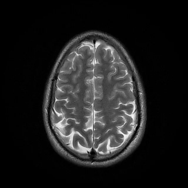
Brain MRI
An MRI (Magnetic Resonance Imaging) is a powerful diagnostic tool that uses a magnetic field and radio waves to create highly detailed images of the brain. It is particularly effective for visualizing soft tissues and can detect subtle changes from a TBI, such as bruising, swelling, and diffuse axonal injury, which might not be visible on other scans. Advanced MRI techniques like DTI (Diffusion Tensor Imaging) can also reveal damage to white matter tracts and neural pathways that may not be visible on conventional MRI scans. This level of detail allows for a precise diagnosis and treatment plan.
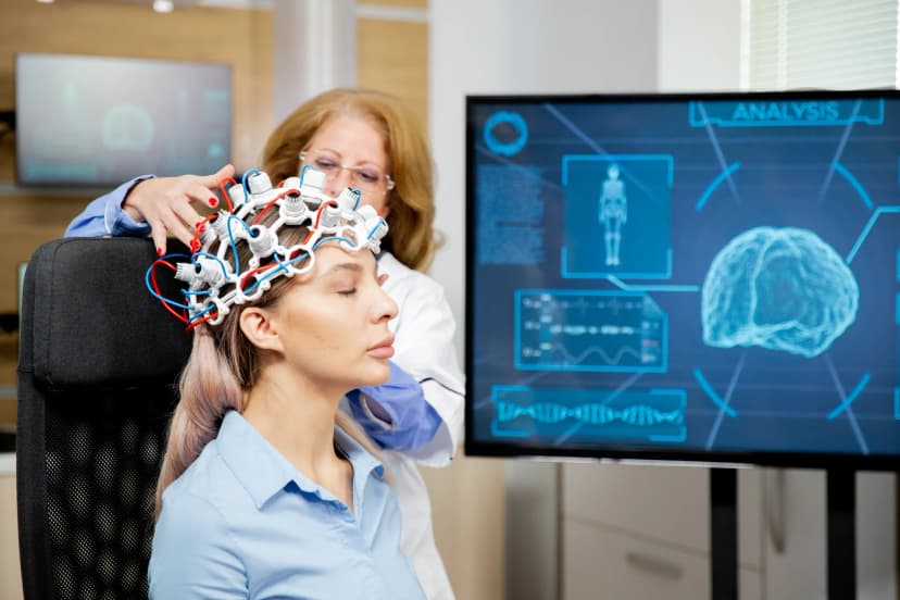
qEEG (Brain Mapping)
A qEEG (Quantitative Electroencephalogram) provides a detailed analysis of brainwave patterns, helping identify the specific location and severity of brain dysfunction. This is a non-invasive diagnostic test that measures electrical activity in the brain. An electrode cap is placed on the scalp to detect and record brain wave patterns. This procedure helps diagnose various neurological conditions including epilepsy, sleep disorders, brain tumors, and brain damage. The test lasts 40 minutes and is completely painless. EEG results can provide valuable insights into brain function and help guide treatment decisions for neurological conditions.
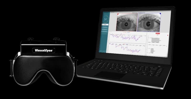
VNG
VNG is a type of diagnostic test that evaluates involuntary eye movement called nystagmus. Nystagmus could point to an inner ear (vestibular) disorder. Symptoms associated with a vestibular disorder include: balance problems, dizziness, lightheadedness, tinnitus (ringing in the ears) and/or vertigo.
1. Ocular testing: The patient watches different dots on a monitor while keeping their head still.
2. Positional testing: The patient is instructed to move their head or body to different positions.
3. Caloric testing: A small amount of warm, cool, and ice cold water is introduced into each ear. These different water temperatures should cause nystagmus.
Our Treatments
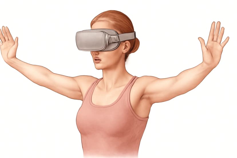
VRT (Vestibular Rehabilitation Therapy)
Traditional vestibular rehabilitation therapy is a type of physical therapy that involves exercises to help manage dizziness and balance issues. VRT is done in our office using Virtual Reality (VR). Our VR modules are a series of computer-generated simulations and games that were specifically designed to help patients reduce symptoms of dizziness, vertigo, and loss of balance.
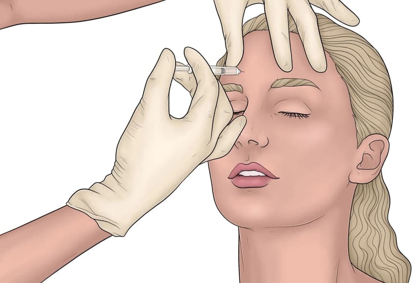
Botox for Posttraumatic Headaches
Botox injections are done to treat posttraumatic headaches by injecting the botulinum neurotoxin in the head and neck muscles to help them "relax" by reducing the muscle contractions which transmit pain signals to the brain that we interpret as headaches. Botox injections are FDA approved to treat headaches.
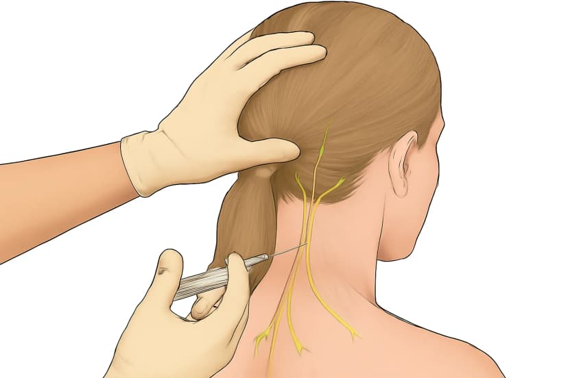
Greater and Lesser Occipital Nerve Block
Occipital nerve block injections numb the greater and lesser occipital nerve to help relieve pain. These nerves can become irritated or compressed, leading to chronic headaches, neck pain, and scalp sensitivity. The injection helps reduce inflammation and blocks pain signals from these nerves.
Treatment Options
Many of our procedures can be performed using either steroid injections or PRP (Platelet-Rich Plasma) therapy, depending on your specific condition and treatment goals.
Steroid Injections
Corticosteroid injections are a common treatment used to provide fast and effective relief from pain caused by inflammation in the spine or joints. Inflammation is often the source of pain when nerves, discs, or joint spaces become irritated and swollen. By delivering medication directly to the affected area, corticosteroid injections help reduce this irritation, allowing the surrounding tissues to calm down and function more normally.
PRP Therapy
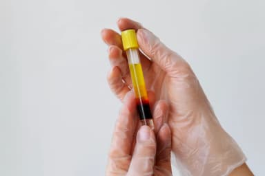
Platelet-Rich Plasma (PRP) therapy is a regenerative treatment that uses your body's own natural healing abilities to repair damaged tissues and reduce pain. The process begins with a small sample of your blood, which is placed in a centrifuge and spun to concentrate the platelets. These platelets are rich in growth factors — powerful proteins that stimulate tissue repair, reduce inflammation, and support healing in joints, ligaments, tendons, and discs. By injecting this concentrated solution directly into the injured or painful area, PRP helps the body accelerate its natural recovery process.